MRI biopsy needles
Detection susceptibility artifact areas are the key ‐ Accurate position for MRI needles
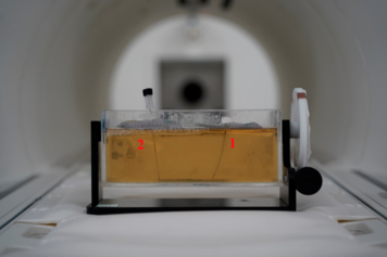
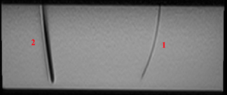
Magnetic resonance imaging (MRI) is used as a guiding tool in minimally invasive procedures. Due to its superior soft-tissue contrast, MRI is the ideal guiding and monitoring system for standard needles. However, one of the main challenges in these procedures is the susceptibility of needles to generate artifacts caused by the interaction of compatible materials with magnetic fields and significantly distort the images. Most of the cases a minimum of fivefold from the original needle’s dimensions, making it extremely difficult to visualize the needle’s shaft and tip. Therefore, the needle placement inside a tumour or a specific area could not be precise and time-consuming for clinicians. As a result, there is a continuous need to use new materials for the needles to optimize their visualization in magnetic resonance imaging. Non-metallic materials can reduce these artifacts. We propose a design of a coaxial needle with fibre enforced inner core and an outer hollow sheet. The concept has been evaluated in the MRI environment as shown in the figures below.

Fig.1. Biopsy needle prototype labeled No. 1 and reference NiTi biopsy needle
labeled No. 2 aligned inside gelatine phantom

Fig. 2. MRI image of needles inside the phantom, needle labeled No. 1 appears
with low artifacts while NiTi needle labeled No. 2 appears as a huge artifact


